Home

Wickeln Shake Folgen hypodensité scanner Erwähnen nimm Medizin Verschlingen

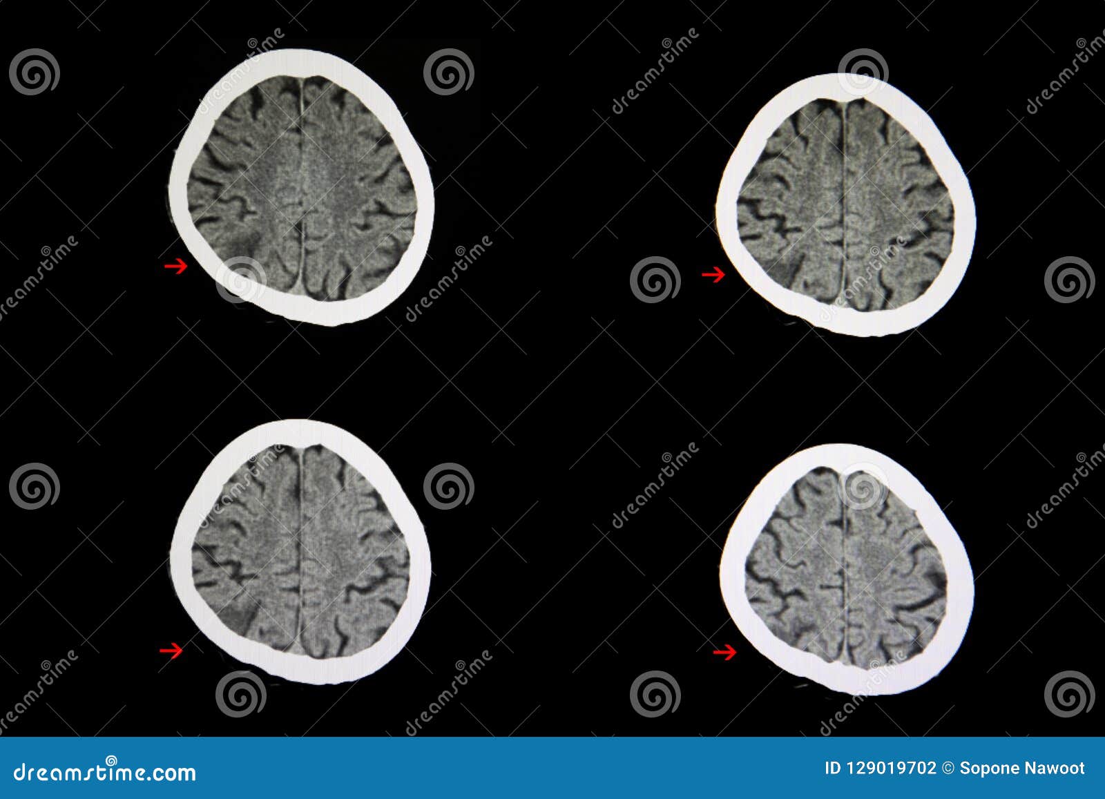
CT Scan Cerebral Infarction Stock Photo - Image of lesion, medicine:  129019702
CT Scan Cerebral Infarction Stock Photo - Image of lesion, medicine: 129019702

cours AVC
cours AVC

View Image
View Image

PPT - Neuroradiologie - Neuroimagerie PowerPoint Presentation, free  download - ID:3526927
PPT - Neuroradiologie - Neuroimagerie PowerPoint Presentation, free download - ID:3526927

Can MS lesions appear hypodense, in white matter, on a CT scan? - Quora

EPOS™
EPOS™

CT brain scan image (A) showing a large hypodense area in the left... |  Download Scientific Diagram
CT brain scan image (A) showing a large hypodense area in the left... | Download Scientific Diagram

PDF] The hypodense artery sign. | Semantic Scholar

Les hypodensités encapsulées : un marqueur scanographique simple pour  prédire la croissance des hémorragies cérébrales et l'issue clinique  défavorable. - Docteur imago
Les hypodensités encapsulées : un marqueur scanographique simple pour prédire la croissance des hémorragies cérébrales et l'issue clinique défavorable. - Docteur imago

The Hypodense Artery Sign | American Journal of Neuroradiology

CT Cerveau: Spectacle AVC Ischémique (hypodensité Au Droit Lobe  Frontal-pariétal) Banque D'Images Et Photos Libres De Droits. Image  31821962.
CT Cerveau: Spectacle AVC Ischémique (hypodensité Au Droit Lobe Frontal-pariétal) Banque D'Images Et Photos Libres De Droits. Image 31821962.

Focal hypodense hepatic lesions on non-enhanced CT (differential) |  Radiology Reference Article | Radiopaedia.org
Focal hypodense hepatic lesions on non-enhanced CT (differential) | Radiology Reference Article | Radiopaedia.org

Recent Advances In... - Recent Advances In Medical Health

High-resolution CT scanning showing a hypodense area over the left... |  Download Scientific Diagram
High-resolution CT scanning showing a hypodense area over the left... | Download Scientific Diagram

CT Head Interpretation | Radiology | Geeky Medics

Une pseudo-leucodystrophie de mauvais pronostic-Scanner cérébral sans  injection. Hypodensité bihémisphérique de la substance blanche  périventriculaire postérieure avec respect du ruban cortical.
Une pseudo-leucodystrophie de mauvais pronostic-Scanner cérébral sans injection. Hypodensité bihémisphérique de la substance blanche périventriculaire postérieure avec respect du ruban cortical.
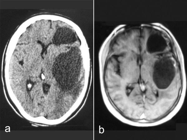
Epileptic seizures caused by encephalomalasic cysts following radiotherapy:  a case report | Cases Journal | Full Text
Epileptic seizures caused by encephalomalasic cysts following radiotherapy: a case report | Cases Journal | Full Text
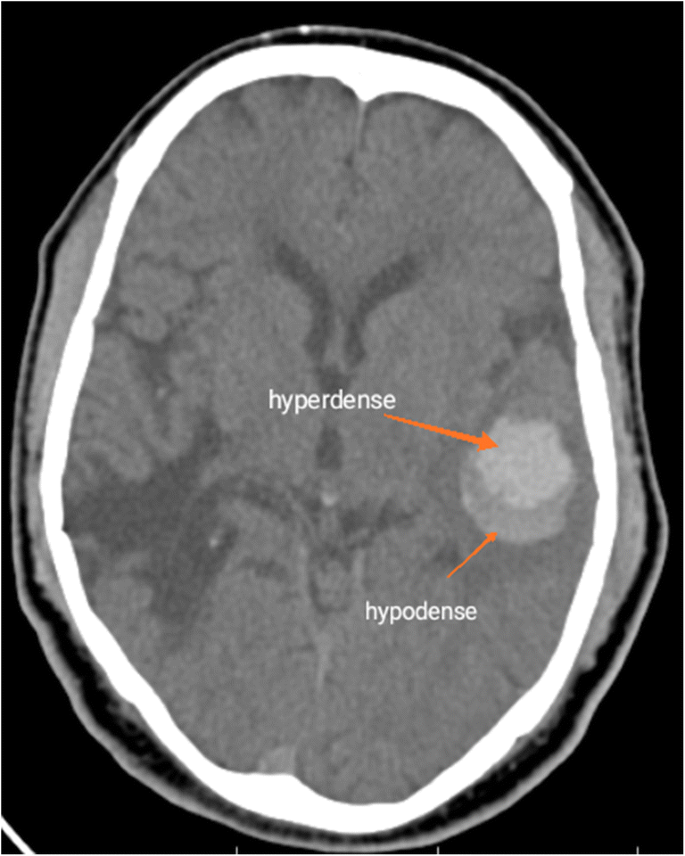
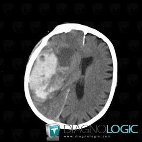
Hypodensité périventriculaire - Diagnologic

Semiologie semio neuroradio

Scanner (CT)
Scanner (CT)

Figure 1 | Cystic Meningioma Simulating Arachnoid Cyst: Report of an  Unusual Case
Figure 1 | Cystic Meningioma Simulating Arachnoid Cyst: Report of an Unusual Case

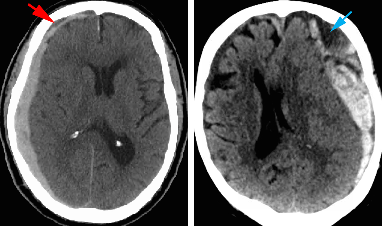
Acute Subdural Hematoma | The Neurosurgical Atlas

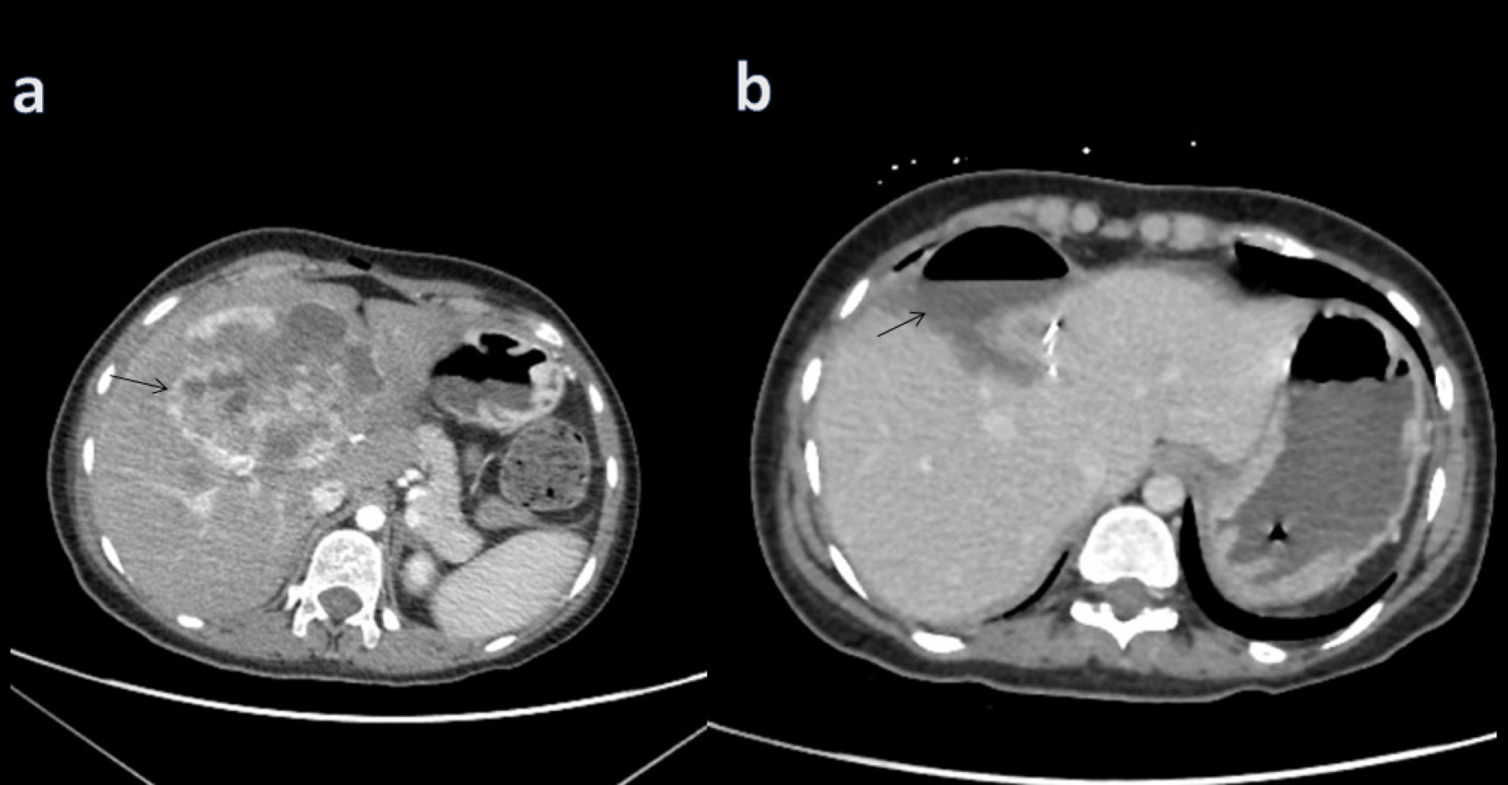
Cureus | Primary Hepatic Neuroendocrine Tumor: A Case Report and Literature  Review
Cureus | Primary Hepatic Neuroendocrine Tumor: A Case Report and Literature Review

Can a Non-Contrast CT Scan of Brain Predict Hematoma Expansion in Acute  Intracerebral Hemorrhage? | SpringerLink
Can a Non-Contrast CT Scan of Brain Predict Hematoma Expansion in Acute Intracerebral Hemorrhage? | SpringerLink